Taking the time to understand the symptoms and risks associated with hematomas is an important step toward protecting your health and well-being.
A hematoma is more than just a bruise. While some are harmless, others, particularly those affecting the brain, can lead to serious health problems if not recognized and treated. Understanding the signs of a hematoma, especially a chronic subdural hematoma, can help you or your loved ones seek medical care when it’s most needed.
At Vascular Neurology of Southern California (VNSC) in Thousand Oaks, Dr. M. Asif Taqi and his team offer specialized neurological care, including the diagnosis and treatment of brain-related hematomas. This article will guide you through what hematomas are, why they occur, and how they are treated.
What Is a Hematoma?
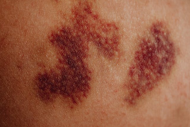
A hematoma forms when blood collects outside a blood vessel, typically following an injury. On the surface, this may look like a bruise, but deeper hematomas—particularly those in the brain—are not always visible.
A subdural hematoma occurs when blood pools between the outer covering of the brain and the brain itself. As blood fills this space, it can increase pressure inside the skull, which may damage delicate brain tissue. While some subdural hematomas develop rapidly, others progress slowly over time and may go unnoticed until symptoms appear.
Common Causes of Hematomas
Most hematomas are caused by trauma, such as a fall, accident, or blow to the head. However, not all cases require major force.
In older adults, for example, the brain naturally shrinks with age, stretching the small veins that cross the space between the brain and skull. Even a minor bump can cause these veins to break and leak blood. Infants are also vulnerable, as their blood vessels are more delicate.
Other factors that increase the risk of hematoma include the use of blood-thinning medications, conditions that affect blood clotting, and repeated head injuries over time.
Symptoms to Watch For
The symptoms of a brain hematoma depend on how much blood has accumulated, how quickly it builds up, and the pressure it places on brain tissue.
Some signs may appear immediately after an injury, while others may develop gradually over days or weeks. Symptoms can include:
- Persistent headaches
- Confusion or difficulty concentrating
- Slurred or slowed speech
- Trouble with balance or walking
- Weakness or numbness in arms or legs
- Changes in vision
- Drowsiness or fatigue
- Nausea or vomiting
- Seizures
In infants, symptoms may include increased head size, irritability, or feeding difficulties.
If you notice these symptoms after a head injury—or even without a clear injury history—it is important to seek medical evaluation.
What Is a Chronic Subdural Hematoma?
A chronic subdural hematoma develops when blood slowly collects under the outer covering of the brain, often over several weeks or months. This typically follows a minor head injury, particularly in older adults whose brains have slightly shrunk over time.
Because the bleeding is slow, symptoms may not appear right away. In some cases, people may not even remember the original injury that caused the problem. By the time symptoms arise, the accumulated blood may already be placing significant pressure on the brain.
When to Seek Medical Attention
Recognizing when to seek medical help is essential. You should consult a doctor if you or someone you care for experiences persistent headaches, changes in mental clarity, speech difficulties, weakness, or difficulty walking after a head injury.
It’s especially important to be cautious if the person is elderly, takes blood-thinning medications, or has a history of repeated falls. Early diagnosis can make a meaningful difference in outcomes.
Diagnosing a Hematoma: What to Expect
At VNSC, evaluation begins with a careful medical history and neurological examination. If a hematoma is suspected, imaging tests are typically used to confirm the diagnosis.
A CT scan is often the first choice, as it can quickly identify areas of bleeding or swelling. In some cases, an MRI may be recommended to provide a more detailed view of the brain. These imaging techniques help determine the location, size, and type of hematoma, guiding the next steps in care.
Treatment Options
Treatment for a chronic subdural hematoma depends on its size and the symptoms it causes. Some small hematomas may be monitored over time with follow-up scans, while larger or symptomatic hematomas often require intervention.
One common treatment is burr hole drainage, where a small opening is made in the skull to allow the trapped blood to drain. For larger collections, a craniotomy may be performed to remove blood clots.
Another approach, middle meningeal artery embolization, uses minimally invasive techniques to block the blood supply feeding the hematoma. This helps reduce bleeding and encourages the hematoma to resolve over time.
In addition, medications may be prescribed to reduce swelling or prevent seizures, depending on the patient’s needs.
Why Choose VNSC for Neurological Care?
The care of patients with chronic subdural hematomas requires specialized knowledge and experience. At VNSC, Dr. M. Asif Taqi and his team are dedicated to providing thoughtful, evidence-based care tailored to each individual’s situation.
With access to advanced diagnostic tools and a range of treatment options, the team at VNSC works closely with patients and their families to ensure that care decisions are made carefully and with attention to long-term health.
Schedule a Consultation
If you or a loved one has experienced a head injury or is showing signs that may suggest a hematoma, consider seeking an evaluation.
Vascular Neurology of Southern California is located in Thousand Oaks and serves patients from West Lake, Woodland Hills, Ventura, Camarillo, Calabasas, and surrounding communities. To schedule a consultation, call (805) 242-4884 or contact us online.